
A crucial safety concern for UHF MRI is the significant RF power deposition in the body in the form of local specific absorption rate (SAR) hotspots, leading to dangerous tissue heating/damage.

The rapid and successful advancement in ultra-high field (UHF) MR scanners (7T or higher) have shown an improvement in the spatial and temporal resolution and the signal-to-noise ratio (SNR) per un
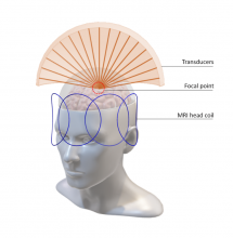
Magnetic resonance guided focused ultrasound (MRgFUS) is a non-invasive therapeutic modality for neurodegenerative diseases that allows real-time imaging of targeted regions.
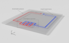
MRI relies on a dense array of radiofrequency (RF) coils to obtain functional and anatomical information inside the body.
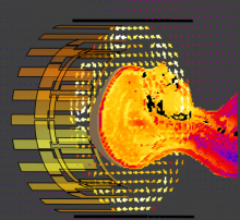
The Winkler Lab has a dedicated radiofrequency (RF) team with a top-notch RF lab to design, demonstrate, and test high field (HF) and ultra-high field (UHF) receive and transmit coils.
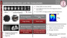
The following set of slides gives an overview of past research topics on UHF MRI at Stanford University:
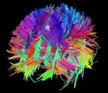
National Institutes of Health (NIH) K99/R00 funded: One of the greatest challenges of modern biomedical science is the mapping of the human brain to understand underlying functionality and behavior


