The rapid and successful advancement in ultra-high field (UHF) MR scanners (7T or higher) have shown an improvement in the spatial and temporal resolution and the signal-to-noise ratio (SNR) per unit time of magnetic resonance (MR) images. However, imaging of human-sized objects at high frequency (Larmor frequency of 1H ≥ 300 MHz) presents a key limitation quantified by an excessive RF power deposition and localized tissue heating due to the shortened in-tissue wavelength. The safety index provided by current MRI scanners is the global specific absorption rate (SAR), which measures the power delivered per mass of tissue, whereas local SAR is generally not given and hard to predict.
To date, existing MR safety-assessment methods based on temperature or electric field measurement are unable to assess the local SAR accurately and non-invasively in vivo.
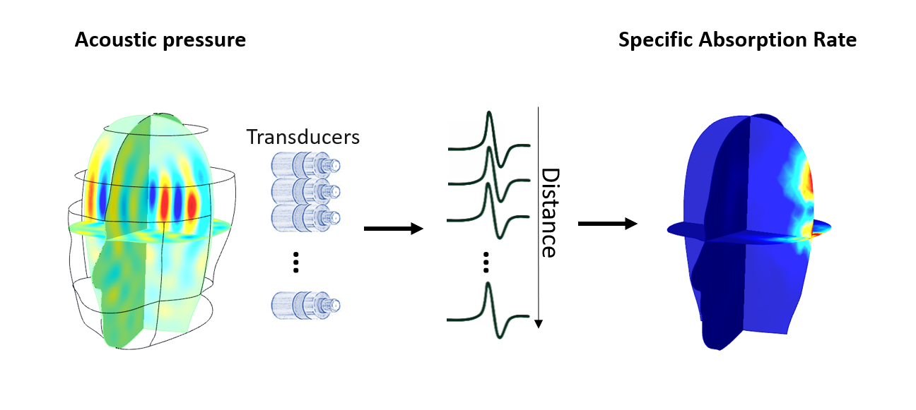
In 2017, Winkler et al proposed a direct SAR monitoring tool based on the thermoacoustic effect. During the RF transmission phase, the absorbed RF energy by the biological tissue increases the local temperature that causes local thermal expansion, resulting in a pressure distribution that propagates through the human body as an acoustic wave.
Our research aims to benefit from the thermoacoustic effect to develop a setup consisting of an array of transducers to predict spatially varying local SAR patterns in vivo at 7T.