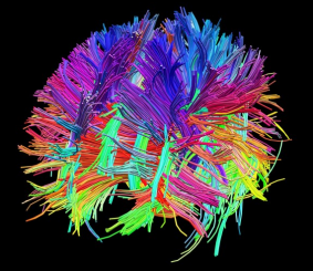
National Institutes of Health (NIH) K99/R00 funded: One of the greatest challenges of modern biomedical science is the mapping of the human brain to understand underlying functionality and behavior. The NIH-funded Human Connectome Project (HCP) is a large-scale, multi-institutional effort that constructs a vast in-vivo database of neural connectivity pathways, acquired using diffusion and functional magnetic resonance imaging (MRI).
This proposal focuses on a key limitation of the HCP that is pivotal for its success: the problem of limited resolution (about 1.5 mm at 3T) that is a critical barrier in the deciphering of structures such as intra-cortical, and very small sub-cortical, network hubs.
Ultra High-Field (UHF) MRI (using magnetic field strengths of 7T and above) as part of the current and future HCP mandate, in combination with high-performance head gradient hardware, offers promise of a crucial improvement over 3T HCP in spatial resolution and sensitivity for deciphering subtle features that are <1 mm in size, and could thus allow mapping of intricate detail.
While the HCP includes UHF field strengths in its mandate, incorporating a plan for 7T connectivity mapping of a population of 200 healthy adults, the bulk of HCP data has been acquired to date at 3T. The reasoning for this: the substantial hurdles that still need to be surmounted before the promised performance increases towards higher sensitivity, and sub-mm resolution can be fully realized. This successful grant application has therefore been focusing on 2 key technological challenges that represent critical barriers holding back UHF brain connectivity mapping:
1) The problem of non-uniform main magnetic field B0, leading to image distortion and artefact, which I am attempting to solve by more advanced “shimming” methods (tools to make the field more uniform).
2) The problem of non-uniform power deposition in the body, which is a safety concern of UHF MRI that has not been completely solved to date. I have proposed an innovative solution involving new technology for direct mapping of power deposition patterns (measured as the specific absorption rate (SAR)) based on thermoacoustic imaging.
My long-term goal is to develop hardware technology for individual patient connectome generation. My approach and primary goal for this 5-year project is to develop a single-device, compact, helmet-style radiofrequency (RF)-coil array, thus facilitating the generation of human connectome maps with sub-mm resolution by overcoming a critical barrier that currently limits the full potential of the HCP.
The proposed research will not only benefit the safety of UHF HCP. It will significantly impact the long-term clinical potential of HCP by creating an individual patient connectome as a future clinical diagnostics tool. This will be accomplished with new approaches to testing and/or identifying abnormal brain circuitry in individual patients. Comparisions will then be made to the standard healthy adult atlas. Intrinsically improved resolution and sensitivity will offer the capacity for a collective extraction of neural circuitry with sufficient detail to provide underlying brain connectivity. This could lead to a near-future standard clinical tool that will not only benefit our understanding of brain connectivity, but will also provide a mainstream tool for diagnosis, monitoring, and treatment guidance for a variety of conditions such as neurodegenerative and psychiatric disorders.